Embryology Biology 441 Spring 2010 Albert Harris
Lecture notes for Monday, January 25, 2010
The parts of the body that are made of ectodermal cells:
Cells that don’t move to the interior during gastrulation are (therefore?!) considered to be ectoderm. It is a good question why decisions about future differentiation of cells are so often made by mechanical rearrangements ("Morphogenetic Movements"). It’s a rather odd way to make such decisions, and it's also odd that it gets ignored by most people inventing theories about the signals that control which embryonic cells will differentiate into which cell types.
The ectodermal cells themselves are then sub-divided into three categories
(#1) Somatic ectoderm
-
Epidermis layer of skin,
and fingernails, hair, feathers, scales (of reptiles & birds)
(teleost scales are mesodermal, however)
Lens of eye,
Inner ear [cochlea etc.] (formed from Otic placode)
Olfactory placode -> Sensory nerves of nose
Other Placodes which form nerves in head area.
Other Placodes that form the Lateral Line of fish & amphibia
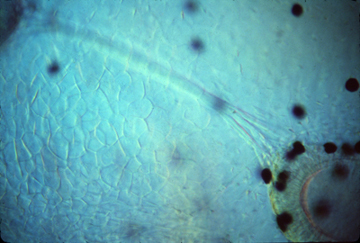
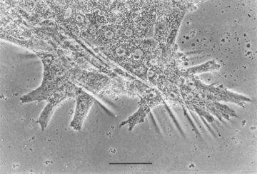
Neuromast cells have special filopodia that detect even very small bending forces, which alter the frequency of nerve impulses sent to the brain. These filopodia are supported by fibers of the muscle protein actin (and are much smaller than hairs on your head, to which they are only similar in shape.
&&&&&&&&&&&&&&&&&&&&&&&&&&&&&&&&&&&&&&&&
While we are on the subject of neuromast cells and the inner ear:
Inertia is detected by water flow in the 3 semi-circular canals bending filopodia of sets of neuromast cells located in parts of these canals.
Gravity is detected by the weight of two (mutually perpendicular) sets off otoliths
which are crystals of calcium carbonate attached to the filopodia of other hair cells.
Sound is detected by long rows of neuromast cells whose filopodia get vibrated against
by special flaps of tissue, arranged so that different frequencies of sound will mostly
stimulate different neuromast cells (because of combinations of different resonances.

In mammals, the sound-detecting part of the inner ear is lengthened and coiled to form a structure named the cochlea. Only mammals have a cochlea, but all vertebrates have part of their inner ears specialized to detect sound vibrations. Frog inner ears tend to be tuned to resonate only to certain frequencies, I have read.
*******************************************************************
Back to subdivisions of the ectoderm:
(#2) Neural tube ectoderm
-
Brain
Spinal cord
segmental motor nerves (nerves that stimulate muscles to contract)
(but NOT post-ganglionic autonomic nerves, which are neural crest)
This origin of this name is that the spine seems to be split in two. But it isn't split; what happens is that they two sides fail to fuse.
Spina bifida often results in partial paralysis & can result in non-formation of the brain. 50% of cases of spina bifida are caused by deficiency in the vitamin named folic acid. (& would not occur if women made sure to get enough of this vitamin early in pregnancy)
An early diagnosis of spina bifida is based on finding certain molecules in the amniotic fluid that are normally found only inside the neural tube (in the neurocoel).
___________________________________________________________________

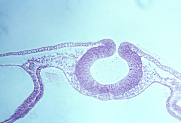
Also formed by out-foldings from the sides of the brain are the optic cups, the outermost edges of which bend back inward to form the neural retina. The part of the optic cup just inward from the neural retina differentiates into a thin epithelium full of melanin pigment granules, called the pigmented retina.
Small areas of somatic ectoderm are induced by the optic cups to fold inward and differentiate into the lens of the eye. Lens cells elongate very much.
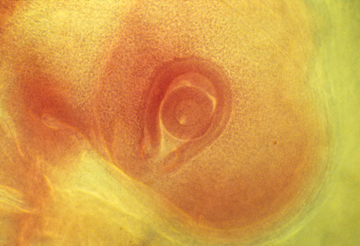
Eye cup with lens
__________________________________________________________________
In the mechanical sense, expansion of vertebrate brains is equivalent to inflation of several closely spaces balloons. Cells pump water into the neurocoel, creating enough water pressure to expand the anterior part of the neural tube. The fore-brain, mid-brain andhind-brain are areas which stretch more than the part of the brain between and behind them. Eventually, there is much more cell growth in these areas; But at first, they are thin-walled, water-inflated & balloon-like
Experiments have shown that the brain won't expand if the fluid pressure is released. The eye-ball also depends on hydraulic expansion for its enlargement. (and won't enlarge if punctured)
_________________________________________________________________
(#3) Neural crest ectoderm (sometimes called "ectomesenchyme")
-
segmental sensory nerves (connected to spinal cord) (NOT optic nerve, etc.)
postganglionic autonomic nerves (including the medulla part of the adrenal gland)
Pigment cells of skin ("melanocytes" in Mammals) (plus amphibia, fish, birds & reptiles also have other pigments than melanin and the cells that made these other pigments also develop from neural crest)

-
Schwann cells (that wrap their plasma membranes around nerve axons
(in the peripheral nervous system, to form the myelin sheath.)
-
Facial skeleton (in the anterior part of the head, the cartilages and [probably?] other tissues that are formed by mesoderm in other parts of the body)
It is a big puzzle why neural crest should form skeletal tissues.
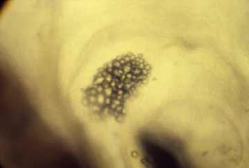
Odontoblasts which are special mesenchymal cells that secrete the inner, dentin layer of the teeth.
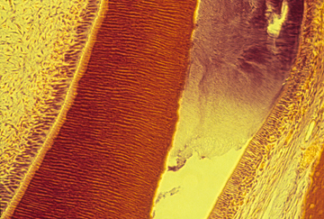
Thin section through tooth development on a mouse embryo.
Enamel is stained purple; dentin is stained dark red
-
#3 ½ ?) Stomodeum (oral cavity cells, also considered to be ectodermal,
rather than endodermal! Although this may have been more an arbitrary decision than a fact based on any particular evidence, I suspect.)
Lining of mouth (and also nasal cavity) (posterior to oro-pharyngeal membrane)
The palate is the roof of your mouth (but not in birds, reptiles, amphibia) The palate is formed by fusion of right and left palatal shelves.
Enamel (outside layer of teeth: secreted by ameloblasts)
Rathke's pocket -> Anterior Pituitary Gland
(That secretes FSH, LH, etc.)
At least one of the three pairs of salivary glands
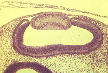
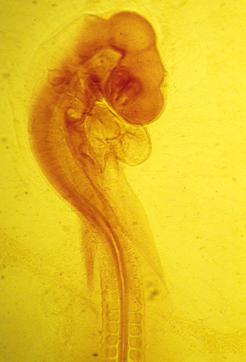
Can you find the lens, neural retina, pigmented retina, otic vesicle, and Rathke's pocket, and other embryonic structures in this photograph..
_____________________________________________________________________
A side comment:
Two secret weapons of the vertebrate subphylum are myelination and
the random DNA splicing clonal selection immune system.
(In case you ever wonder why squids aren't running the planet!) [or are they?]
These are two special advantages vertebrates have over all other animals.
______________________________________________________________________
Review questions, that you should be able to answer on future exams:
a) Are all sensory nerves derived from the neural crest? What about nerves from the nose, eye and inner ear?
b) Are all motor nerves derived from the neural tube? What is a big exception?
c) Are all autonomic nerves derived from neural crest? (what about pre-ganglionic nerves)
d) Do all vertebrate have lateral line systems? What are some that do? & don't?
e) How are placodes related to the somatic ectoderm?
f) Is skin ectodermal or mesodermal? (what about the thickest, heaviest, strongest parts)
g) Why do the early stages of development of the inner ear look so much like the lens?
h) What is an example of a placode that retains an open connection to the outside surface.
i) What is the similarity (at the causal level) between these three birth defects "Spina Bifida", "Cleft Palate", "Cleft Lip" (= so called "Hare Lip")
j) There are human genetic syndromes that combine some or all of the following symptoms: defective tooth dentine; defective autonomic nerves; irregular skin pigmentation, facial skeletal abnormalities, defective adrenal gland. Deduce a possible single underlying cause, based on embryological cell origins.
k) What are the three main subdivisions of the ectoderm? (and name at least two organs that develop from each)
l) If you were in a debate club, and assigned to argue in favor of the idea that there are "really" 4 or 5 subdivisions of the ectoderm, then what would these 5 be? And what arguments could you make in favor of them being as different as the 3?
m) Suggest arguments (pro or con) that the semi-circular canals must have evolved originally as parts of the lateral line system that became separated from the rest of this system, and entirely internalized.
n) Suggest further arguments that the sound frequency-detecting and gravity-detecting parts of the inner ear probably evolved from the semi-circular canals.
o) Think of as many examples as you can in which sharp folds in epithelial cell sheets become boundaries between cells that differentiate into different cell types.
p) Imagine that you are an embryonic ectodermal cell who aspires to differentiate into some kind of a nerve cell: If you were located in the wrong place to have become part of the neural tube, then do you have any further chances of differentiating into a nerve? (List as many further opportunities as you can)
q) * Do all neural tube cells become nerve cells, anyway?
r) * Are myelin sheaths formed by Schwann cells crawling around and around nerve axons, or is there another possibility? What about in the formation of myelin sheaths formed by oligodendrocytes?