The second hour exam will focus on the following subjects:
#1) How physical and behavioral properties cause anatomical shapes.
#2) Development of mesodermal and endoderm organs (and also structures that develop from the stomodeum, proctodeum and pharyngeal clefts).
#3) Cleavage and gastrulation of teleost fish embryos.
#4) Extra-embryonic membranes of Reptiles, Birds and Mammals. Extra-embryonic membranes of Teleost fish.
#5) A little about twinning, especially monozygotic and conjoined twins.
**********************************************************************
Of these, the most important is:
How physical and behavioral properties cause anatomical shapes.
Properties include pressures, strengths of contractility, strengths of adhesion, crawling, turning, frequencies and directions of turning, and probably you will be able to think of some others.
For example, Steinberg's theory depends on a pulling force being exerted by the process of forming cell adhesions, like water is pulled into contact with paper and many other materials.
Remember the computer simulations of moving dots that turn at certain frequencies based on changes or differences of concentration of chemicals diffusing from the cursor (the mouse arrow). You ought to be prepared to draw your own graphs of probabilities of turning, or directions of turning, or speed of movement, based on changes of distances, or differences of distances, of each point to the cursor.
For example, draw three different graphs that correspond to three different ways of causing dots (cells) to accumulate at higher concentrations near wherever the cursor currently is. Also, you ought to be able to draw graphs of behaviors that would cause cells to run away from the cursor, and get as far from it as they can (as if pushed).
Also, you ought to be able to interpret changes in surface curvatures (of embryos, of the heart, of blood vessels, of lobes of the brain, etc.) in terms of strengths of tensions at surfaces and pressures pushing across surfaces or other interfaces (flexible boundaries).
If the exam has drawings of a series of curved surfaces, you ought to be able to figure out the combinations of tensions and pressures that can produce the shapes and changes of shape would produce these shapes (and shape changes) shown to you in drawings.
For example, in the video on Zebrafish gastrulation some interesting changes in surface curvatures occur.
You should be able to deduce relative tensions in surfaces and interfaces, and when these tensions get stronger and weaker at different locations, as the embryo develops
You also ought to be able to figure out how to use adhesion gradients to confirm or disprove Steinberg's Differential Adhesion Hypothesis, according to which cells are pulled to locations of greatest adhesiveness, specifically by the force of attraction toward cell surfaces (this is called the "reversible work of adhesion", and exists between some surfaces, but not between others. An alternative explanation is whether the cells are pulling actively outward along their edges, in a tug of war, so that cells accumulate where cell-substratum adhesions are strongest (or break least frequently).
What can you learn from these two videos:
Can you distinguish whether cells are pulled by forces of attraction between cell surfaces and other objects ("Reversible Work of Adhesion"), or whether cells pull themselves by active pulling forces (traction) along their surfaces. If the latter is true, can you distinguish whether pulling is stronger at parts of the cells touching more adhesive materials, as compared with preferentially breaking adhesions connecting cells to less adhesive surfaces. In all cases, explain your reasoning. Is there a real difference between the edge of a cell being pulled more strongly, versus less frequent breakage of adhesions along that edge of a cell? Is the net result the same? By what criteria can you distinguish between these alternatives? Can you invent third or fourth possibilities? Would any of your answers depend on whether each cell had many leading edges, pointed in different directions, as opposed to having a single leading edge, pointed in only one direction at any one time, but capable of turning?
Suppose we had a chemical attached to a surface that stimulated detachment of cells from their adhesions: How would cells behave on a concentration gradient of such a substance? Would your answer depend on whether each cell had many leading edges, pointed in different directions, as compared with having a single leading edge, pointed in only one direction at any one time, but capable of turning?
And how could you improve the use of such surfaces, with any pattern of differing adhesiveness that you can invent, so as to help prove, or disprove, the Differential Adhesion Hypothesis.
Incidentally, in that time lapse video of the fish egg developing, how would the changes in shapes and surface curvature be explained (interpreted) based on the beliefs of the Differential Adhesion Hypothesis? (Hint: Would increased work of adhesion between adjacent cells cause surfaces of the embryo to bulge outward, or cause it the bulge less? What about bulging that occurs before before the embryo has cleaved into many different cells, or in areas which have not cleaved, or never cleave.
Look at the following video
and try to distinguish what forces are causing sponge cell aggregates to attach, fuse and round up.
Are some parts of their exposed surfaces contracting more strongly than others? (If so, which parts?)
Are cells at the aggregate surface crawling actively into the interior? (How can you tell?)
Are cells being pulled passively into the interior by Reversible Work of Adhesion (pulling force exerted by maximization of cell-cell adhesiveness)? (How can you tell?)
Because cell contractility is weaker where cells are touching other cells on all sides, but stronger where cells are in contact with the surrounding water? (How can you tell?)
Design experiments (=imagine and create situations) which maximize the expected differences in what happens, depending on whether fusing of aggregates, rounding up etc. are caused by reversible work of adhesion, active cell contractility, weakening of cell contractility where cells contact each other, or other theories.
For example, what if you put the masses of cells on adhesion gradients, or put them in contact with flexible rubber, added a soluble chemical that weakened cell contractility (or one that strengthened contractility), a chemical that weakened or strengthened cell-cell adhesiveness. What alternative behaviors can you imagine should happen?
For example, what if you spread a layer of dissociated sponge cells onto a rubber sheet, and the cells contracted the rubber and caused visible wrinkles. Next, suppose that you dropped spherical cell aggregates (like the ones in the video) onto the upper surface of the layer of sponge cells that is wrinkling the rubber.
The possible results are:
-
A) No change in the wrinkling of the rubber.
B) Increase in the wrinkling of the rubber.
C) Decrease in the wrinkling of the rubber.
This is a time lapse video of living sponge cells inside a living sponge (not dissociated).
Can you figure out what combinations of cell behaviors are creating and remodeling the canal systems through which sponges suck water? Having seen what goes on inside a sponge that isn't dissociated, does it change your opinion about whether Wilson was correct about "archeocytes" (= stem cells).
As an example of theories about forces predicting shape changes, and specifically about designing experiments for which opposing theories predict opposite results, please consider the following illustration from a very good textbook in cell biology:
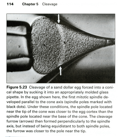
Ray Rappaport's classic "Doughnut Experiment" had proved that cleavage furrows are induced by overlap of signals from the poles of mitotic spindles, and not by signals from the chromosomes, or anything else located at the metaphase plate half-way between spindle poles.
This led to the questions
ONE) Whether the signals from the poles induce weakening of cortical contraction (with the furrow forming where this weakening is least, or
TWO) Whether the signal from the poles induces strengthening of cortical contraction (with the furrow forming where the strengthening is greatest)
In this experiment, you can see that a cell has been squeezed into a conical shape, and that the cleavage furrow has formed nearer the narrower end of the cell.
Can you figure out whether the results of the experiment (that is, the shifting of the location of furrow induction nearer the thinner end of the cell) support theory ONE and contradict theory TWO?
Or do the results support theory TWO and contradict theory ONE?
Or neither?
Into what other shapes could you distort a cell, in order that the effect on furrow location would tell you the most about whether furrows are induced by stimulatory or inhibitory signals?
What does the caption imply?
Why or how was this experiment designed, would you guess?
What might you be able to prove or disprove by distorting spherical cell aggregates into different shapes?
Do you happen to know how Newton figured out that gravity decreases in strength in inverse proportion to the square of distance? Did he, maybe have a really tall ladder, and carried a weighing scale up to higher altitudes?
Imagine gravity decreased inversely with the first power of distance (instead of the square), then how would this change the shape of the moon's orbit, the orbits of planets.
If you had a magic power to change the law of gravity (for example, changing the exponent in the inverse law of gravity), then could you steer planets into different orbits without having to push or pull on them?
What do you think of that as an analogy for the methods by which genes create and maintain anatomical shapes?