Lecture notes for Wednesday, April 6, 2016
Limb bud development
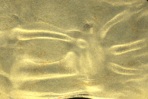
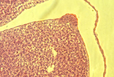
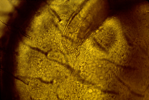
Photo: Cartilages differentiating inside a living chicken hind-limb bud
Limb buds develop into legs, arms, wings, or fins.
Normally, embryos form 4 limb buds,
2 anterior & 2 posterior.
But are there ways to induce salamander embryos to form three legs on each side?
(Like the Martians in "John Carter of Mars"!)
Surgically implanting otic or olfactory placodes, or plastic beads containing Fibroblast Growth Factor, will cause three legs to form on the same side.
Scientists suspect that embryos re-use the same inductive signals in different parts of the body; and inductively competent is already concentrated along both flanks.
Limb buds develop if transplanted to other parts of the body, and to the amnion of chicken embryos.
You can also split limb buds, fuse limb buds, rotate them upside down, or backwards.
If you cut an early limb bud in two, it can form 2 arms, or often it will form a branched leg (mirror images).
Fusing two limb buds can often form only one leg. (surgically graft one limb bud right next to another one, & they merge! These kinds of experiments work best in salamander embryos.)
Notice the similarity to what Driesch discovered! That splitting early echinoderm embryos in two results in each half developing into a half-sized "scale model" embryo.
Several body organs have been discovered to form two organs, for example two complete hearts.
Another example is eyes, and also the nose rudiment, which can be either split into 2, or two can be fused into one.
The human uterus also forms by side to side fusion of the lower end of the oviduct. (Muellerian Duct).
[Be prepared for an exam question: Describe at least ? embryonic organs that develop by fusion of two masses of cells. List examples of organs or organisms being split into two, where each half is able to form all parts normally developed when they are not split.]
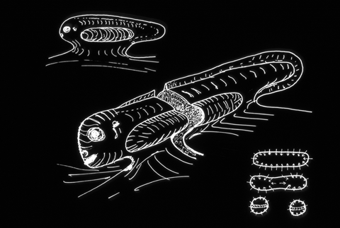
Some kinds of parasitic worms often split limb buds in frogs, resulting in 5 or 6 hind limbs!
&&&&&&&&&&&&&&&&&&&&&&&&&&&&&&&&&&&&&&&&&&&&&&&&&&&&&&&&&&&&&&&
Development of the 3 geometric axes are controlled separately in limb buds And at 3 different times, by 3 different sets of chemical signals
-
Proximo-distal axis
Anterior-posterior axis
Dorso-ventral axis
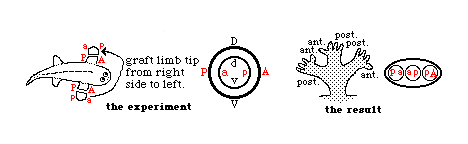
During a later period of development, limb buds are no longer able to regulate (change) their anterior-posterior axis, but can still regulate the dorso-ventral axis!
In effect, that means that the part that would have developed into the palm of the hand can change its fate, and develop the geometry appropriate to the back of the hand, and vice versa, but a rotated limb bud can no longer change where the thumb forms, etc.
Analogous surgical experiments were done with the inner ear (otic placode) cutting it out and grafting it back in, upside down and backwards, etc.
Before a certain stage, it "regulates" completely, in the sense that the different cells change how they develop according to their new orientations, such that the semi-circular canals, otoliths, etc. all develop at locations that are normal with respect to the rest of the body.
At a later stage of development, the dorso-ventral and medio-lateral axes are still able to regulate, but the anterior-posterior axis is irreversible.
These rotation experiments were done in salamanders, in the 1910s-30s.
(the limb bud experiments and also the inner ear experiments, and many others.)
&&&&&&&&&&&&&&&&&&&&&&&&&&&&&&&&&&&&&&&&&&&&&&&&&&&&&&&&&&&&&&&
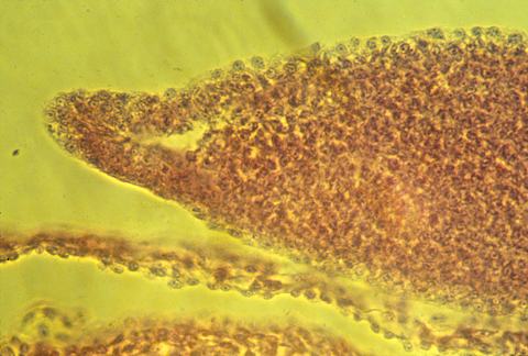
The Apical Ectodermal Ridge (AER) is a thickening of the epidermis
that forms along the outer rim of the limb bud
(of bird, mammal, reptile,
frog and fish embryos - but NOT on limb buds of salamanders!)
In frog embryos, the AER is comparatively small and indistinct, and for many years, everyone believed that frogs didn't have an AER. But more careful studies found them. The scientific meeting at which this evidence was first presented was in Wales in 1971, & I was in the audience!
Surgical removal of the AER causes failure of the distal structures to develop. Cut the AER off early, then no elbow, forearm, wrist or hand. Cut the AER off later, then no wrist & hand.
This was discovered by Prof. John Saunders, who taught for many years at the State University of New York in Albany, and who visited this department a few years ago, and is one of the most stimulating scientists I have ever met.
In sections of bird AERs, the epithelial cells seem to be pinched together at their basal ends, and the ridge is as much a "pucker" as a thickening.
Sections through mouse embryo AERs show a different geometry. It is definitely a thickening, but with some indication of contraction at Both basal and apical surfaces.
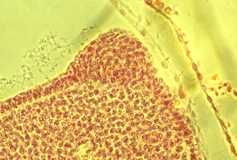
Here is a photograph of an oblique, or slanted section through a chicken AER.
The developing tail fins of fish have a similar thickened ridge.
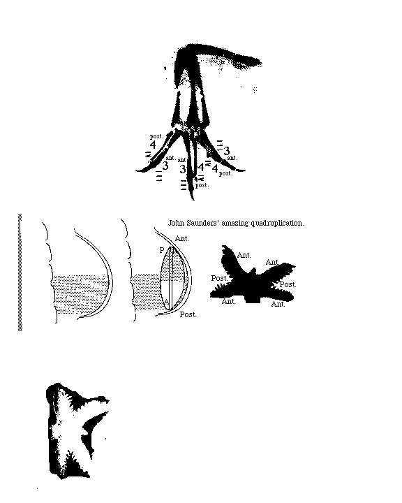
Grafting an extra AER can cause a double wing to form. A mutation in chickens causes double AERs and double wings!
But the AER doesn't itself become any these structures.
It somehow signals to them to form.
There has been much research and several theories on this subject.
Fibroblast growth factor 10 seems to be the key signal molecule.
FGF10 can replace the effect of the AER;
If you cut off the AER, & put a bead coated with FGF in its place
then the distal limb structures will form more or less normally.
(If anybody has done this with snake embryos, I haven't heard about it!
Probably, snakes still have the genes for legs, and maybe the signal receptors.)
Legless lizards and legless salamanders might also be subject to such experiments.
Another interesting fact is that salamanders (especially newts) are the only kinds of vertebrates that can regenerate cut off legs (more on this in the next lecture), and also are the only kind of vertebrate that don't form an AER.
However, during regeneration of salamander legs, an epithelial thickening is formed at the tip of the regenerating limb stump! This thickening is not elongated, however.
I suggest to you that some big conceptual breakthrough is waiting for you to discover!
If the AER cells are a special shape, does their shape mean they are more contractile, or what? Or is it an evolutionary remnant of fish fin shapes?
The proximo-distal axis is considered to be controlled by the AER.
The anterior-posterior axis can be reversed by grafting cells from the back side of the limb bud to the front side, which is called the "Zone of Polarizing Activity" (ZPA)
What is the chemical basis of this? Is it retinoic acid? Mirror-image branching legs can be produced by putting retinoic acid onto the anterior side of an early limb bud.
And if you put RA on the regenerating tail stump of a frog, then one or several legs will grow instead of a new tail!
And putting RA on the regenerating leg stump of a salamander will cause the entire proximo-distal sequence of structures to be regenerated, even if only the hand or wrist had been cut off!
The newer evidence is that the sonic hedgehog protein controls the anterior-posterior axis formation.
The dorso-ventral axis of limb development: Can be reversed by removing the entire limb bud ectoderm and turning it upside down. The key protein is thought to be one of the Wnt proteins.
Organization of fingers somehow depends on certain hox genes: Specifically hoxD 9, 10, 11, 12, 13
The spaces between fingers are made by programmed cell death = "apoptosis" activation of cytoplasmic enzymes, that digest cells from the inside out.
The webbing of a duck's foot results from a lack of apoptosis.
And by the way, many cancer chemotherapy drugs actually work by stimulating apoptosis, rather than by direct harm to the cancer cells. The drugs were intended to kill fast growing cells, but don't really work that way!