This is an excerpt from notes for Biology 441, Vertebrate Embryology.
Somites were mentioned in the Biology 446 class on October 26th and perhaps in some others. Only some of the slides below were actually shown. The main point for the concept of shape homeostasis is that spatial regularity is part of what we mean by shape.
Spatially-periodic structures whose locations are caused by where somites form:
Segmental structures that are ectodermal:
-
Dorsal root ganglia (aggregations of neural crest cells that differentiate into sensory nerves)
Motor nerve bundles
"Dermatomes" in the medical sense. Areas of skin that are innervated by each different segmental sensory nerve.
(I have gotten the impression that motor nerve innervation patterns also are restricted
to analogous bands, like dermatomes)
Somite Formation
At first, there are continuous columns of "paraxial" mesoderm.
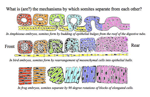
The drawing above shows the geometrical rearrangements that some scientists have reported to cause somite segmentation in different classes of chordates.
Top: Amphioxus, Middle: bird and mammal, Bottom: frogs. Anterior is to right side
Then these columns spontaneously split apart, one pair at a time.
As if a quonset hut were to split into a row of igloos.
Or as if a long piece of french bread were to slice itself.
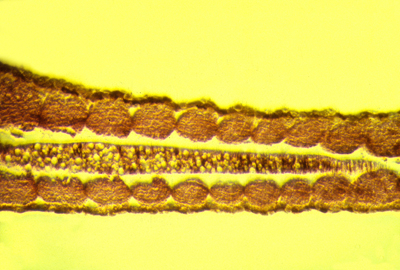
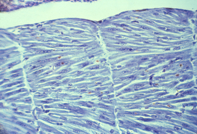
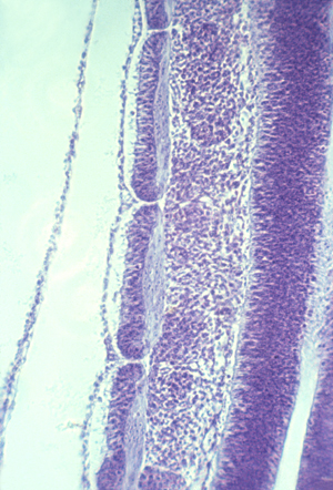
Longitudinal section of notochord and somites in a developing frog embryo
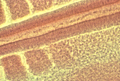
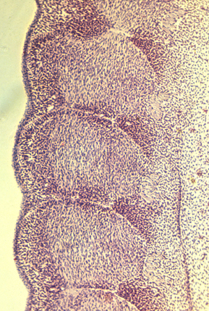
Somites forming in a fixed whole mount chicken embryo. Anterior to left.
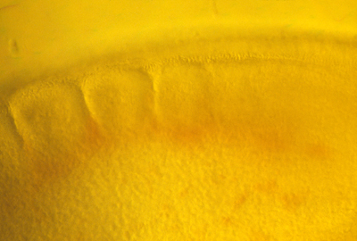
Somites forming in a living chicken embryo. Anterior to left.
The physical mechanism of splitting apart has now been discovered to be stimulation of decreased cell-cell adhesions by specific binding of ephrin proteins.
Ephrins stimulate cells to break their adhesions to each other, and guide optic nerve fibers to their map-like patterns of connections in the roof of the mid-brain.
Some textbooks say that ephrins cause cell to repel each other, but really they induce detachment of adhesions: Cell traction pulls each side in a tug of war, therefore weakening of cell adhesions causes them to crawl directionally away from the weakened adhesions.
In scanning EM photos, "somitomeres" can be seen, where a somite is going to form, apparently as the first stages of somite formation.
In most species, somites start as hollow epithelial balls, and conversion of cells from being mesenchymal to epithelial therefore seems to be part or the process of separation. (These epithelial cells seem to have basal surfaces outward.)
For most species, the same number of somites forms in each embryo.
(the number is 32-34 in humans, 50 in chicks, 65 in mice (many in tail),
more than 500 in some snakes, and varies with temperature in fish!
You form as many vertebrae as your embryo had formed pairs of somites.
Each somite subdivides into four parts:
The dermatome --> cells form the inner layer of skin (dermis)
The myotome ---> all the skeletal muscle cells of the body
The anterior sclerotome --> the posterior half of a vertebra!
The posterior sclerotome --> the anterior half of a vertebra!
Not a typographical error! Each somite gives cells to 2 vertebrae
And each vertebra develops from cells of two adjacent somites.
That way muscles run from one vertebra to the next,
and motor and sensory nerves come out between vertebrae.

Somites only temporarily exist in the embryo:
but their spacing controls future segmentation of many tissues;
vertebrae, ribs, arterial branches
and also sensory ganglia (that develop from neural crest!!)
and also motor nerve bundles from the spinal cord.
Incidentally, to Neurologists a "dermatome" means the stripe-shaped areas of skin and muscles that are innervated by sensory and motor nerves from each segmental spinal nerve. In the disease "Shingles", stripe-shaped zones of blisters form on the skin (looking like shingles on a roof) because rows of blisters form on the skin of different dermatomes.
Neurologists have a method for mapping paralysis caused by slipped disks, by inserting electrodes under the skin and firing one set of nerves at a time. This method hurts quite a bit.
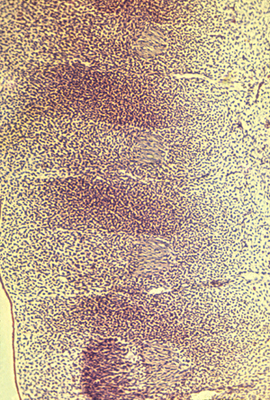
Three somites:
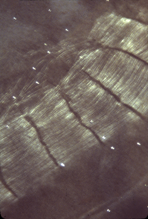
Myotomes in a salamander
Polarization microscopy shows that myotomes develop muscle fibers very early.
Alternating anterior and posterior sclerotomes
Nerve ganglia form only in the anterior sclerotomes. Also notice that small arteries run just below (behind?) each of these ganglia.
Although somites somehow cause the locations of these nerve ganglia and the arteries branching from the aorta,
neither the nerves or arteries develop from somite cells.
Surgical transplantations of salamander somites (5 from the tail replacing 3 from the trunk) prove
that anatomical segmentation is caused by somites themselves,
NOT as additional effects of what-ever signals cause somites to form.
Longitudinal section of 3 somites, that goes right through the myotome
Notice the bundles of nerve fibers located just to the right (toward the midline of the embryo) of each of these three myotomes.
Myotomes right under them
Then sclerotomes
The continuous blue stripe on the right is the neural tube.