Review for the third hour exam, part one
1) In the development of the heart, do we first form a one-chambered embryonic heart, then replace it with a two chambered fetal heart, which we use to pump blood while we construct the 4-chambered heart that we will use after birth and for the rest of our life?(Hint: no) But what do we do instead?
2) In what way is that imaginary sequence of hearts analogous to what actually happens in the embryonic development of kidneys?
3) A serious birth defect that sometimes occurs is that a hole connects the right and left ventricles of the heart. This is analogous to what other birth defects in other? Hint: It isn't called "cardia bifida" but that might have been chosen as a name.
(Does it result from something having cut or cleaved a hole through the septum between the ventricles?)
Hint: no
4) Until birth, blood flow and oxygenation of blood is not abnormal in babies with holes connecting their two ventricles. Why not? Please explain why oxygenation of blood then suddenly become very abnormal in these babies, at the time of birth (but worked OK before birth).
5) Do mammal embryos ever develop a hole connecting their right and left atria? (Trick question!)
Yes, a hole always forms - and what shape is this window-like hole? Oval shape of foramen ovale
6) Write the order of blood flow through an embryo at 5 weeks starting with blood in the peripheral circulation. Terms that may or may not be used: Inferior Vena Cava, Sinus Venous, Aorta, Truncus Arteriosus, Foramen Ovale, Primitive Ventricle, Right Ventricle, Left Ventricle, Ventricular Septal Defect, Primitive atria, Right Atrium, Left Atrium, Pulmonary Trunk (artery), Ductus arteriosus, Umbilical Artery, Umbilical vein, Bulbus Cordis.
ANSWER: The key is that this is an embryo at 5 weeks, so certain structures haven't developed yet. We still have a primitive heart tube. Thus the order is Sinus Venosus -> Primitive Atria -> Primitive Ventricles -> Bulbus Cordis -> Truncus Arteriosus
7) From which germ layer is the heart derived? A. Ectoderm B. Mesoderm C. Endoderm D. Cardioderm?
ANSWER: B. Mesoderm
8) Compare and contrast an Atrial Septal Defect and a Patent Foramen Ovale
ANSWER: Both are "holes" between the right and left atria, leading to a shunt. An ASD is due to an error with missing tissue during atrial septation; whereas, a PFO is simply the failure of the foramen ovale to fuse and close after birth.
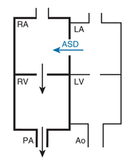
Here is an example of a box diagram for an atrial septal defect. The bold boxes mean fluid overload.
--------------
10) Compare or contrast the three kinds of explanations that scientists have advocated as for sorting out of dissociated cells. (including Malcolm Steinberg's theromodynamic explanation.)
[hint: are there logical similarities or equivalences in the theories, and in the kinds of evidence that are needed to prove which explanation is correct?]
11) If you dissociate a neural tube into its individual cells, and then randomly mix these with dissociated cells from the "somatic ectoderm" (i.e. the cells that will form the outer layer of the skin), then the neural tube cells will sort out to the internal position.
Argue pro and/or con whether this means that the cause of cell sorting must have the same underlying mechanism as the cause of normal neurulation.
Remember that embryos of mammals, salamanders, and frogs form their neural tubes by folding of a sheet of ectodermal cells, and sealing the edges together, leaving a fluid-filled space inside, called the neurocoel. In contrast, embryos of teleost fish form their neural tube from a solid rod of cells, which somehow hollows itself out down the middle (that is, instead of folding and sealing the edges).
Argue pro and/or con whether this means that teleost neurulation has a different causal mechanism than the neurulation of mammal embryos, frog embryos and salamander embryos.
12) Briefly describe and/or discuss whether your opinion or conclusions on the questions above are changed by each of the following facts:
-
a) When sorting-out occurs between neural tube cells and somatic ectoderm cells (in any vertebrate), the mass of aggregated neural tube cells hollows out to form a neurocoel-like cavity.
b) The anterior 80% of a bird embryo's neural tube forms by active folding and sealing of the edges, just as occurs in the whole neural tube of mammal and amphibian embryos; but the rear-most 20% of a bird embryo's neural tube forms by hollowing out of what starts out as a solid rod of cells.
c) If a solid ball of aggregated neural tube cells is put in tissue culture media, next to and in contact with a solid ball of aggregated somatic ectodermal cells, then the latter will extend around the outer surface of the mass of cells derived from the neural tube, until it forms a continuous surface layer. (i.e. the same arrangement as gets produced when these two kinds of cells sort out).
14) According to material posted on the web page, which particular scientist reached this conclusion while stopped at a traffic light?
15) How would (and did) Malcolm Steinberg interpret the ability of dissociated and randomly-mixed cell types to sort out from each other, regardless of whether any given pair of cell types ever normally come in contact in normal development?
16) Because normal animal development never consists of formation of 5 or 6 concentric spheres of differentiated cells, layered around each other, therefore would you argue that the causal mechanism of cell sorting can't be the same as (or over-lap with?) the causal mechanism of normal development?
17) Because no kind of animal develops from an embryo consisting of a random mixture of all kinds of differentiated cell types, which then sort out so as to create anatomical structures, would you say that therefore the mechanism of cell sorting can't be the same as the mechanisms of normal embryonic development?
18) Because mixtures of differentiated cells often rearrange by means of several different sequences of geometrical intermediates, on their way to form the same eventual anatomical shapes, that tells us what? More than one of these conclusions might be true.
Which are true?
1) That different causes can form the same end result?
2) That the same causes can produce different intermediates?
3) That cell sorting is caused by lack of counter-balance between opposed forces?
4) That these forces eventually become balanced?
5) That there are many arrangements in which opposed forces are imbalanced?
6) That (in such a case) there was just one balance in which forces are stably counter-balanced?
7) That the forces producing the rearrangement must all be thermodynamically reversible?
8) That only reversible forces can produce the same end result from different beginnings?
9) That different kinds of cell-cell adhesion proteins must cause sorting out.
10) That different total amounts of adhesion proteins (regardless of kind) must cause sorting out.
Which are true and can be logically deduced from the observed ability of cells to reach the same eventual results by two or more different sequences of intermediates?
Which are not true?
Which are not true, but believed by many scientists?
Which could or would be true, if some particular additional fact were observed or otherwise known?