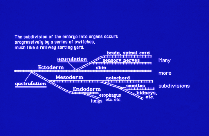
The patterns of germ layer separation from each other, and subdivision into sub-partsFor historical (as well as factual) reasons embryologists say there are three primary germ layers (Keim-Blätter = "tissue-leaves")
Mesoderm Endoderm
2) Upper layer of lateral plate mesoderm 3) Lower layer of lateral plate mesoderm 4) Endoderm
Endo-mesoderm
Just memorize Ectoderm, Mesoderm, Endoderm. (And realize that early embryologists could just as well have chosen a first subdivision into TWO, and called the two Ectoderm, and Endo-mesoderm. Ectoderm subdivides into three parts: (That is an actual fact, not some arbitrary decision)
Neural crest ectoderm --- sensory nerves, skin pigment, Schwann cells, & certain others Neural tube ectoderm ---- Brain, spinal cord, retina of eye, motor nerves & a few others. That is a little joke for the Latin students; don't worry about it. Plus the stomodeum, which is classified as ectoderm.
The process of subdivision of ectoderm is called neurulation (in vertebrate embryos, not urchins). (And "Others" above always means certain specific other organs and tissues, that are the same in humans as in frogs as in chickens. Somewhat different in insects, worms, etc. But even those do have their own kinds of ectoderm subdivisions. Please understand that animal embryos are very conservative (in a good sense) about these subdivisions. It is as if somehow it means a lot to the causal mechanisms that control gene expression. Maybe all genes that are transcribed in ectodermal tissues have some specific DNA sequences in their promoter regions, or something else fundamental like that. Nobody knows; You may be the person who invents, and then experimentally confirms the answer. The words ectoderm and endoderm have also been used by Huxley (Darwin's friend & supporter) as names for the outer and inner epithelia of Hydra, and other Cnideria.
Implicitly, what does that suggest? Nobody knows how to think about evolution of a new differentiated cell type (yet!)
There must have been some molecular basis for evolution of each cell type. Mesoderm subdivides itself into the following (in embryos of vertebrates)
(it doesn't subdivide into parts, like somites, etc.) (Many anatomy textbooks say intervertebral discs develop from notochord) Notochords are sheets of collagen fibers wrapped tightly around vacuolated cells. Intervertebral discs have sheets of collagen tightly wrapped around cartilage . Somites which subdivide into 3 parts Intermediate mesoderm which subdivide... Lateral plate mesoderm which subdivide...
Somites form as pairs (~32 pairs in humans) right and left of the notochord. This separation of somite cells controls how many backbone bones your body will form. For example, if embryos of a given species of snake, fish, bird, mammal forms 46 pairs of somites, then their body will develop 46 vertebrae. (And surgical grafting can change this number!) They would also form 46 pairs of motor nerves, 46 sensory nerve ganglia, 46 branches from aorta. (Not merely tissues that develop from somites, or even that develop from mesoderm. Also, somites are temporary structures; they segment, and then their cells redistribute. but some ghost of where they were remains and controls where nerves, bones etc. develop. Also not yet understood is/are the mechanism(s) that cause splitting of somites from each other. "The Clock and Wave-Front" theory has been very popular, on and off, for many years. There is supporting evidence! But please forgive me if I am not yet convinced. "Oscillator and gradient theory" is another name this could have been called. ----------------------------------------------------------------------------------------------- Each somite subdivides itself into three parts (Scott Gilbert's textbook raised this to 4 )
Myotome: All of the skeletal cells of the body develop from myotome. Even in arms, fingers, … Sclerotome: the cells of which become cartilage and bone. (But much cartilage and bone also develops from lateral plate mesoderm, and also from neural crest! Particularly in the head region.
Intermediate mesoderm forms kidneys, kidney ducts, and sperm ducts. (NOT egg ducts) Anterior intermediate mesoderm becomes pronephros, and pronephric duct.
|
