Lecture notes for Monday, January 22, 2018
Anatomical Shape Formation:
"Morphogenetic movements"
Cells actively move and rearrange to build anatomical structures.Sometimes certain cells merely differentiate at their proper positions, and just stay there.
At the other extreme, the eventual positions of some other kinds of cells have little or nothing to do with where they will eventually be.
Four important kinds of active cell rearrangements that form anatomy:
#1) Invagination (= folding of epithelial cell sheets).#2) Cell crawling ("Amoeboid cell locomotion", Exertion of traction)
#3) Osmotic pressure, Electroosmotic Pressure (the kind exerted by cartilage)
#4) Inflation of water-filled cavities (such as the brain and eye-balls)
-------------------------------------------------------------------------------------------------
Videos shown in this lecture:
a leucocyte (white blood cell) moving by amoeboid locomotion among red blood cellsgastrulation in a sea urchin embryo
neurulation in an amphibian embryo
simulation of neurulation in a chick embryo
Serial sections of a chick embryo were photographed in order from the tail toward the head and assembled into a movie. Folding of the neural tube begins at the head end, so the assembled video is equivalent to looking at the sequential changes of neurulation at a single location.
mesenchymal cells forming clumps as they pull on a collagen substratum
birefringence of collagen viewed through polarizing filters as it is contracted by cells exerting tractional forces
When it's dark, there is no alignment of the collagen. When it becomes bright, this means that the collagen has been aligned.
mesenchymal cells on a silicone rubber substratum, forming wrinkles in the rubber by tractional pulling
Can you figure out where the tractional forces are being exerted?
nerve axons extending by tractional force exerted at their tips
Unfortunately these forces are too weak to wrinkle the rubber.
Also look at the following videos, which were not shown in the lecture:
animation and video of particles moving on the surface of mesenchymal cells
Amoeboid locomotion?
There are many kinds of amoebae; and they crawl by fundamentally-different mechanisms.
Maybe a dozen different means by which cells move from place to place.
Most of these mechanisms depend on actin and myosin.
(But nematode sperm don't even have any actin).
"rolling" amoeboid locomotion, in which the plasma membrane moves in the pattern of a tank tread.
Compare to the movement of the leucocyte
cytoplasmic flow in Amoeba proteus
-----
A video of the embryonic formation of kidney tubules was also shown. This isn't my video, so I can't post it on the web site.
-------------------------------------------------------------------------------------------------
Examples of epithelial invagination (infolding)
The (1) archenteron of sea urchin gastrulas.The (2) neural tube of all vertebrates (except NOT in teleost fish)
Infoldings that become (3) lenses of the eyes,
that become (4) the inner ears.
Infoldings of the endoderm that become (5) salivary glands,
(6) pancreas,
(7) liver,
(8) kidney tubules,
(9) mouth,
(10) nose,
and (11) teeth are formed by invaginations of epithelial cell sheets.
Sample Questions:
A) What are ten examples of organs of the body that are formed by epithelial invaginations?B) Guess how sea urchin embryos form their coelomic cavities?
C) Guess how hair follicles develop.
D) Guess how the neural retina becomes separate from the pigmented retina.
Epithelial folding is believed to be caused by contraction of cytoplasmic actin and myosin when they somehow get concentrated in the concave surfaces of epithelia. (Other mechanisms have been proposed, some based of differential swelling)
Evidence in favor of this mechanism:
Actin staining with a mushroom toxin that selectively binds to actin.
Actomyosin fibers can be seen in transmission electron microscope sections.
Poisons that interfere with formation of actin fibers also inhibit invagination.
More evidence is needed:
Methods to map locations of force, strengths and directions of contractile forces.
Possibilities: Why does one part of an epithelium fold, but another doesn't?
(1) More actin? (2) More myosin? (3) more of both actin and myosin.
And/or actomyosin might become (4) more concentrated at one surface of the cells,
or maybe (5) there is more directionality to their contraction?
What are some other possibilities? By what experiments could you learn more?
-------------------------------------------------------------------------------------------------
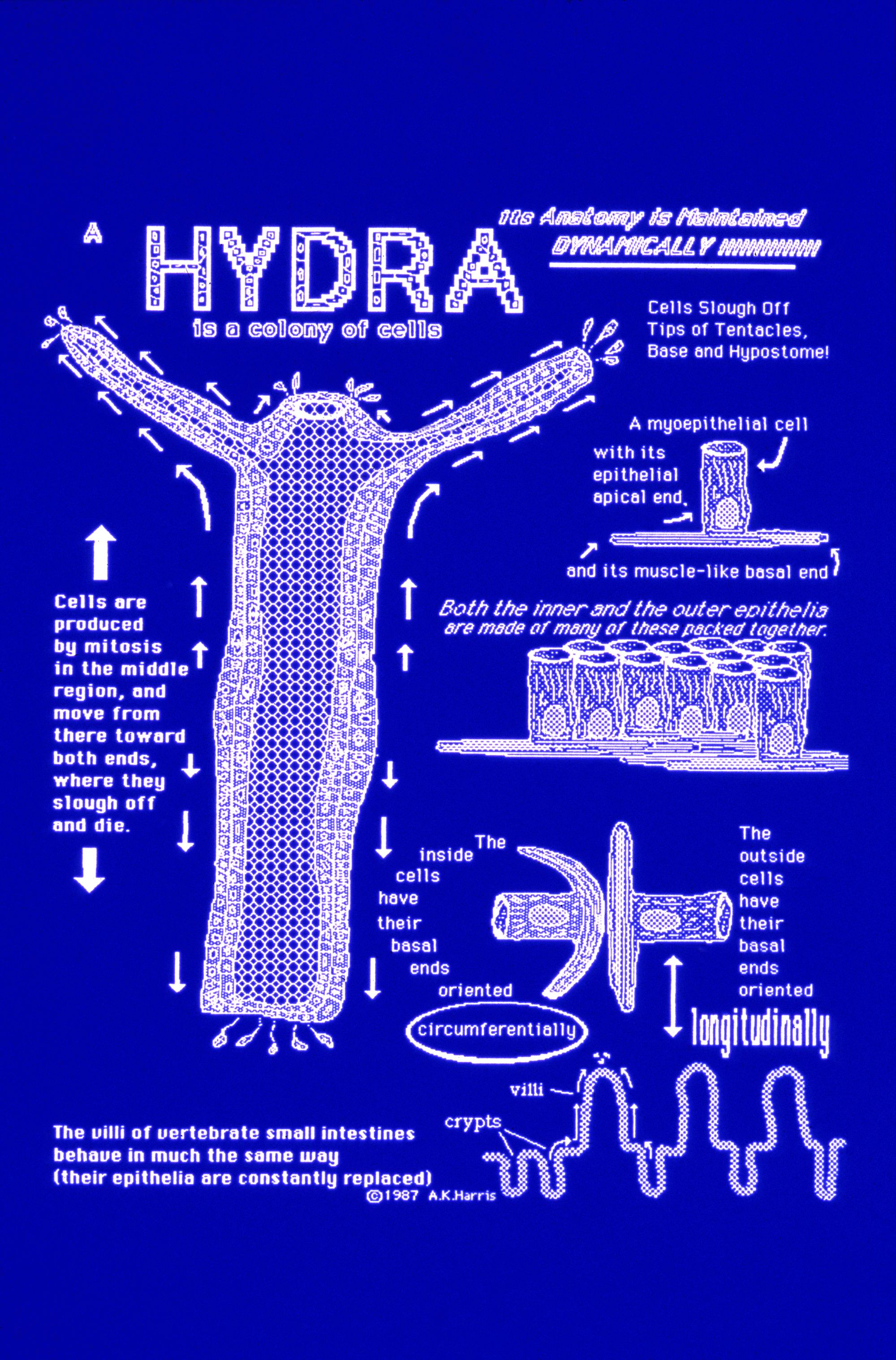
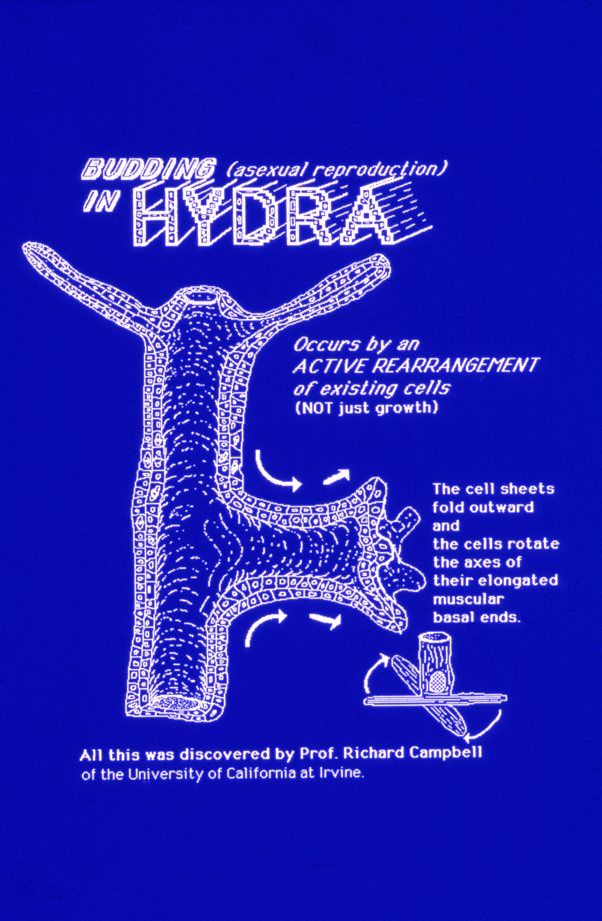
A side issue: How do Hydra cells arrange themselves into hollow cylinders?
Nobody knows! Nobody even knows how to find out! (Perhaps by contracting with greater force in the circumferential direction than in the longitudinal direction?)
Until such questions can be answered for animals as simple as Hydra, what chance is there the problem can be answered for cylindrically-arranged human cells? (Blood vessels, ducts, glands etc.)